Diagnosing Esthesioneuroblastoma
Esthesioneuroblastoma, also known as olfactory neuroblastoma (ONB), is a rare cancer that develops in the upper nasal cavity. Because of its location and subtle early symptoms, diagnosing this tumor can be challenging.
This article will explore the various steps involved in diagnosing esthesioneuroblastoma, including symptom evaluation, and imaging tests, which help guide treatment decisions. Understanding the diagnostic process can help patients and caregivers feel more prepared and empowered throughout this journey.
The Diagnostic Process for Esthesioneuroblastoma
Diagnosis starts with a detailed evaluation of symptoms and medical history. Symptoms can be like those of common sinus issues due to the tumor’s location.
- Common Symptoms: Persistent nasal congestion, frequent nosebleeds, loss of smell, or facial pain. Some may experience vision changes, headaches, or neck swelling if the tumor has spread to nearby lymph nodes. Since these symptoms overlap with other conditions, doctors need to carefully assess their duration and severity.
- Physical Examination: Physicians will inspect the nasal passages, and order further tests if any abnormal growths are found.
Imaging Studies
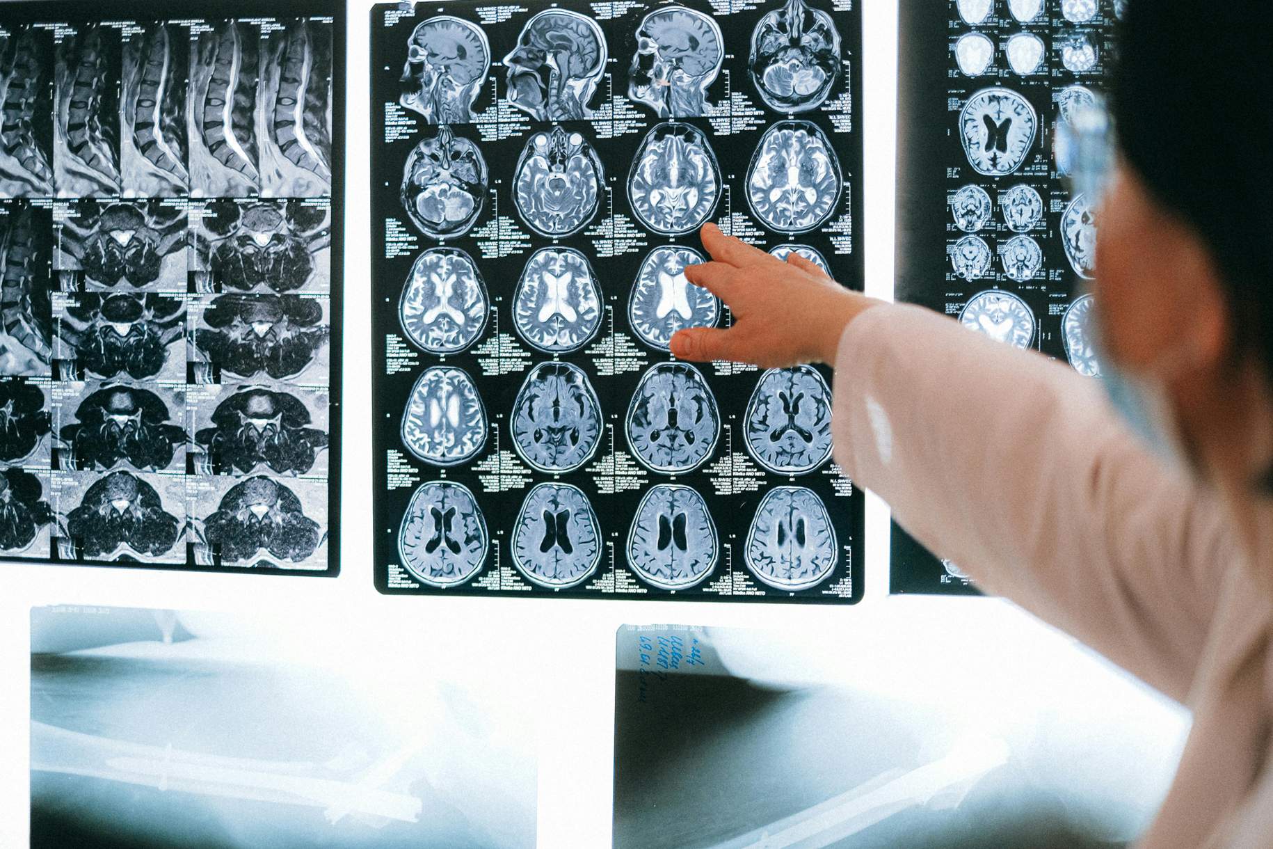
Imaging is a critical part of diagnosis, as it allows doctors to visualize the tumor and determine its size, location, and extent. The most common imaging techniques include:
- Magnetic Resonance Imaging (MRI): MRI is the preferred imaging method as it provides detailed pictures of the soft tissues in the nasal cavity and surrounding areas. MRI can also help determine whether the tumor has spread to nearby structures, such as the eyes or brain.
- Computed Tomography (CT) Scan: CT scans are often used with MRI to provide a clearer picture of the tumor’s space near the bones in the skull, which is useful for surgical planning.
- Positron Emission Tomography (PET) Scan: PET scans may be used to check if the cancer has spread, and also to help distinguish between malignant and benign growths.
Why should you have your surgery with Dr. Cohen?
Dr. Cohen
- 7,000+ specialized surgeries performed by your chosen surgeon
- More personalized care
- Extensive experience = higher success rate and quicker recovery times
Major Health Centers
- No control over choosing the surgeon caring for you
- One-size-fits-all care
- Less specialization
For more reasons, please click here.
Nasal Endoscopy
During this procedure, a thin, flexible tube with a light and camera is inserted through the nostrils, providing a clear view of the inside of the nasal passages. This procedure helps identify abnormal growths, tissue inflammation, or obstructions that may suggest the presence of esthesioneuroblastoma. If any suspicious areas are found, doctors may take a tissue sample (biopsy) during the same procedure.
Biopsy and Pathological Examination
A biopsy is the way to confirm a diagnosis of esthesioneuroblastoma. During this procedure, a small sample of the tumor is removed and examined under a microscope to confirm the presence of cancer cells.
- Types of Biopsies: Depending on the tumor's location, a biopsy can be performed during a nasal endoscopy or through a more invasive surgical approach.
- Pathological Analysis: The tissue is analyzed by a pathologist to determine the type of tumor cells present by looking for specific cellular changes that indicate esthesioneuroblastoma. Further tests can identify markers that help distinguish esthesioneuroblastoma from other types of nasal and sinus cancers.
Tumor Grading and Staging
Understanding the tumor’s grade and stage is essential for developing a successful treatment plan. This provides information on how aggressive the tumor is and whether it has spread to other parts of the body.
- Grading: The tumor is graded based on how abnormal the cancer cells look under a microscope. Lower-grade tumors (Grade 1) tend to grow more slowly, while higher-grade tumors (Grade 4) are more aggressive and have a higher risk of spreading. The tumor grade can impact treatment decisions and help predict outcomes.
- Staging the Tumor: Staging describes the extent of cancer, including how large it is and whether it has spread to nearby tissues or distant organs. The Kadish staging system is often used for esthesioneuroblastoma, with three main stages:
- Stage A: Tumor confined to the nasal cavity.
- Stage B: Tumor extending to the paranasal sinuses.
- Stage C: Tumor involving areas beyond the sinuses, including the orbit, brain, or distant organs.
Key Takeaways
- Persistent nasal congestion, nosebleeds, loss of smell, and other related symptoms often lead to further diagnostic testing for esthesioneuroblastoma.
- MRI, CT, and PET scans are used to visualize the tumor, assess its size, and determine if it has spread.
- A biopsy is essential for confirming the diagnosis, and tissue analysis helps identify the specific type and grade of the tumor.
- These provide important information about how aggressive the cancer is and how far it has spread, helping guide treatment decisions.


